Imaging Anatomy of the Human Brain – 2016
پروسه چاپ این کتاب بین 5 الی 7 روز کاری میباشد
اطلاعات بیشترقیمت منصفانه
ارسال سریع
تنوع و کیفیت بالا
پشتیبانی و پاسخگویی
978-1936287741
ویراست اول
1
رحلی
464
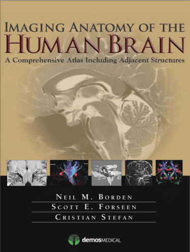
Imaging Anatomy of the Human Brain – 2016
A Comprehensive Atlas Including Adjacent Structures
کتاب تصویربرداری آناتومی مغز انسان – ۲۰۱۶
به راستی یک “اطلس قرن بیست و یکم” ، این مرجع دیداری جامع ، مروری کامل از آناتومی مغزی حاصل از استفاده از روشهای تصویربرداری متعدد از جمله تکنیک های پیشرفته را ارائه می دهد که امکان تجسم ساختارها را با MRI یا CT معمولی امکان پذیر نمی کند.
An Atlas for the 21st Century
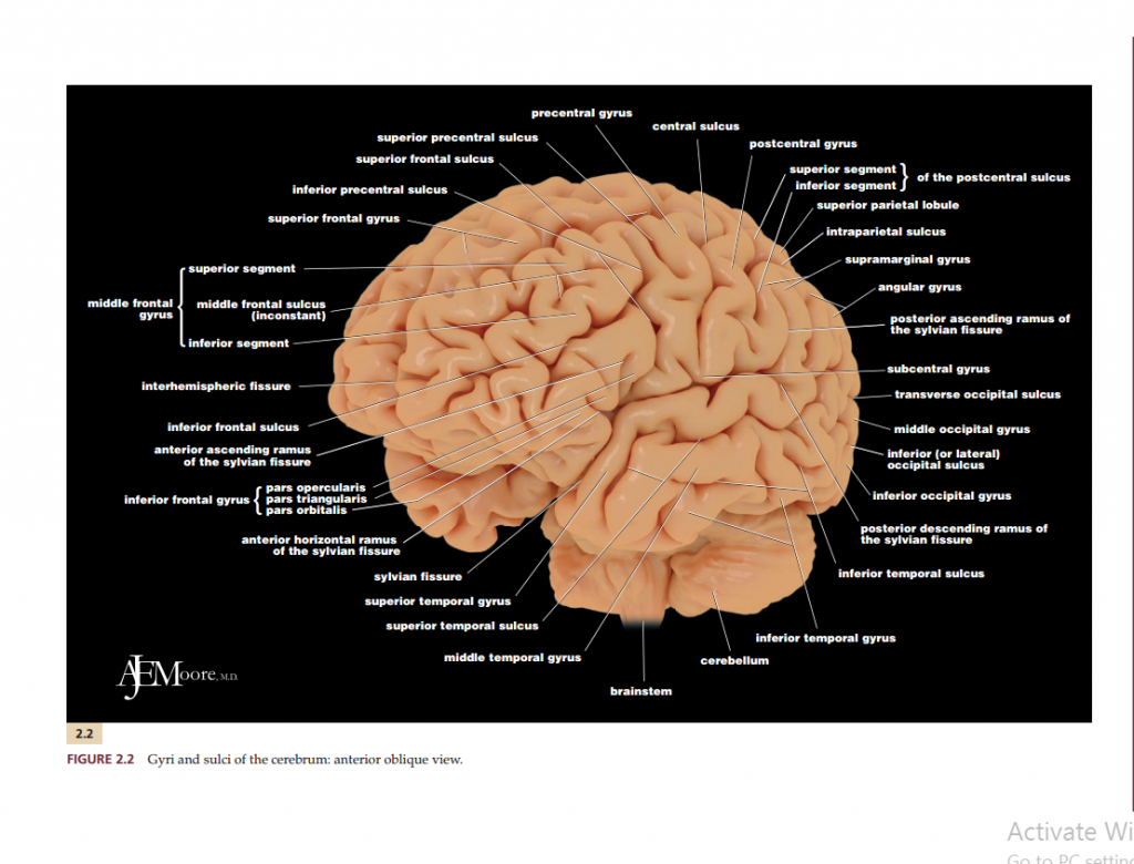
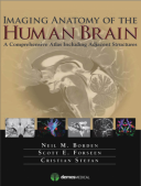
The most precise, cutting-edge images of normal cerebral anatomy available today are the centerpiece of this spectacular atlas for clinicians, trainees, and students in the neurologically-based medical and non-medical specialties. Truly an “atlas for the 21st century,” this comprehensive visual reference presents a detailed overview of cerebral anatomy acquired through the use of multiple imaging modalities including advanced techniques that allow visualization of structures not possible with conventional MRI or CT. Beautiful color illustrations using 3-D modeling techniques based upon 3D MR volume data sets further enhances understanding of cerebral anatomy and spatial relationships. The anatomy in these color illustrations mirror the black and white anatomic MR images presented in this atlas.
Written by two neuroradiologists and an anatomist who are also prominent educators, along with more than a dozen contributors, the atlas begins with a brief introduction to the development, organization, and function of the human brain. What follows is more than 1,000 meticulously presented and labelled images acquired with the full complement of standard and advanced modalities currently used to visualize the human brain and adjacent structures, including MRI, CT, diffusion tensor imaging (DTI) with tractography, functional MRI, CTA, CTV, MRA, MRV, conventional 2-D catheter angiography, 3-D rotational catheter angiography, MR spectroscopy, and ultrasound of the neonatal brain.
by
Product details :
- Hardcover: ۴۶۴ pages
- Publisher: Demos Medical; 1 edition (August 25, 2015)
- Language: English
- ISBN-10: ۱۹۳۶۲۸۷۷۴۹
- ISBN-13: ۹۷۸-۱۹۳۶۲۸۷۷۴۱
- Amazon Best Sellers Rank: #۲,۴۳۱,۴۰۴ in Books (See Top 100 in Books)
- #۲۹۹۶ in Neuroscience (Books)
- #۳۷۳ in Medical Atlases (Books)
- #۱۹۴۱ in Radiology (Books)













دیدگاهها
هیچ دیدگاهی برای این محصول نوشته نشده است.