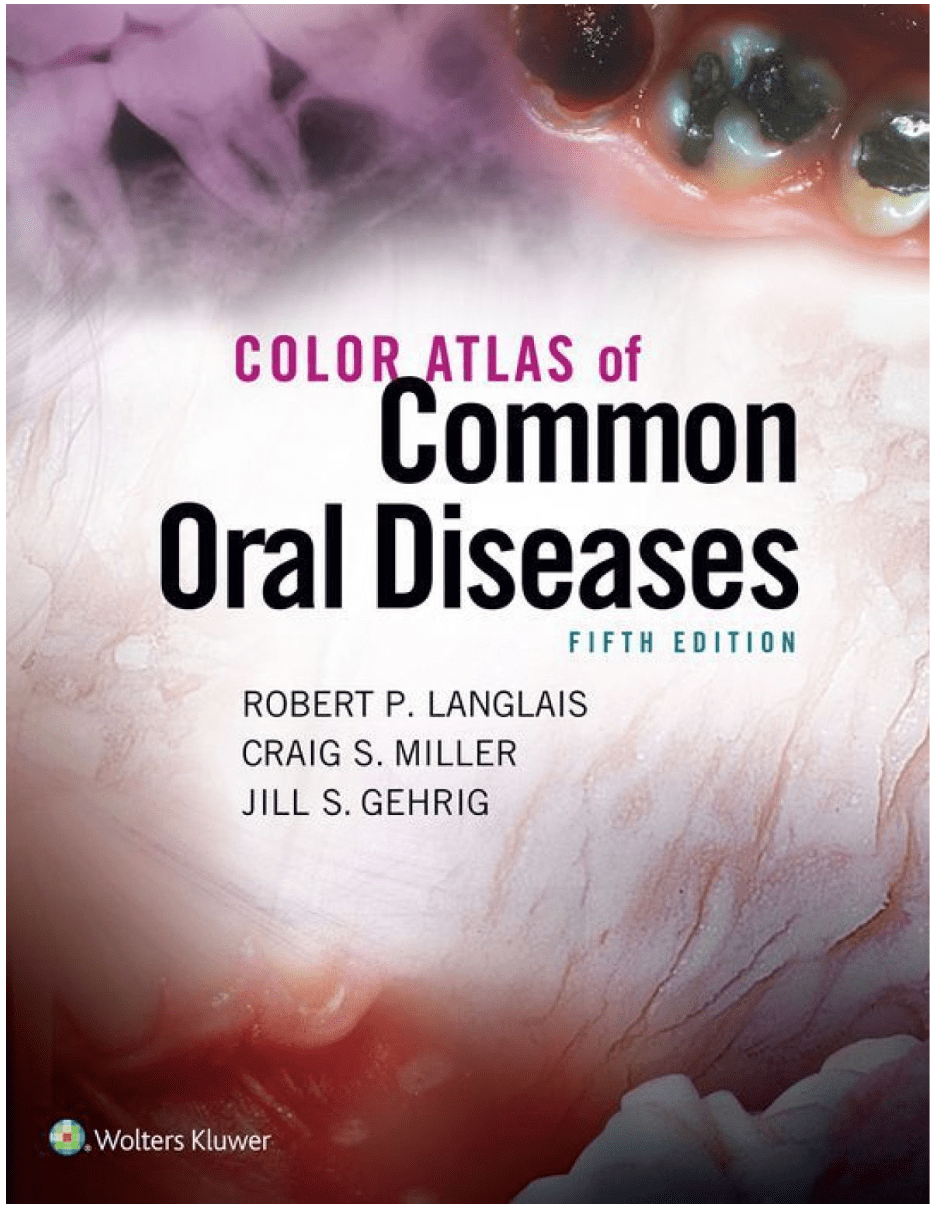
Color Atlas of Common Oral Diseases 2017
رحلی
پروسه چاپ این کتاب بین 5 الی 7 روز کاری میباشد
اطلاعات بیشترقیمت منصفانه
ارسال سریع
تنوع و کیفیت بالا
پشتیبانی و پاسخگویی
9781496332083
ویراست اول
2017
رحلی
288
Color Atlas of Common Oral Diseases 2017
Featuring over 800 clear, high-quality photographs and radiographic illustrations, this fully updated 5th Edition of Color Atlas of Common Oral Diseases is designed throughout to help readers recognize and identify oral manifestations of local or systemic diseases. The new edition includes expanded and updated content and is enhanced by new images, new case studies, a stronger focus on national board exam prep, and more.
The book’s easy-to-navigate, easy-to-learn-from standard format consists of two-page spreads that provide a narrative overview on one page with color illustrations on the facing page. To integrate oral diagnosis, medicine, pathology, and radiology, the overviews emphasize the clinical description of oral lesions, cover the nature of various disease processes, and provide a brief discussion of cause and treatment options.
Features:
An increased focus on national exam preparation is reflected in end-of-chapter questions, entities, cases, and images.
Differential Diagnosis Tables added to each section help students distinguish a particular disease or condition from others that have a similar appearance.
Coverage of new entities, such as Radiopaque Lesions of the Jaw, have been added.
Additional content on Facial & Neck Lesions has been added.
A greatly expanded Radiographs section includes 30 new images and up-to-date content.
Approximately 85 new images enhance the book’s unparalleled photo and illustration program.
New Student Resources: Case Studies (five per chapter) expand students’ opportunities to apply what they’ve learned to clinical situations. Select Disease Fact Sheets and an image bank.
A reader-friendly organization presents clinical and radio-graphic features of common diseases found in the oral cavity according to location, color, surface change, and radio-graphic appearance.
Chapter-opening learning objectives help students focus their studies by setting forth what they need to know after completing the chapter.
Color-coded sections with tabs organize information into logical sections to help students locate information quickly.
Highlighted key words draw student attention to important concepts.
Case studies(80 in all) give students an opportunity to apply their knowledge to clinical situations and prepare for the National Board Examinations.
A comprehensive Glossary offers clear, simple definitions for key terms.
Expanded instructors’ resources and all-new student resources save instructors time and help students succeed.








دیدگاهها
هیچ دیدگاهی برای این محصول نوشته نشده است.